Low Serum Iron: Causes, Symptoms, and Solutions
Introduction
Iron is an essential trace mineral that plays a pivotal role in oxygen transport, cellular energy production, and DNA synthesis. When serum iron concentrations fall below optimal levels, the body’s ability to produce hemoglobin and support metabolic processes is compromised, leading to a cascade of clinical manifestations commonly grouped under the umbrella of iron‑deficiency anemia. This article provides an evidence‑based, in‑depth look at low serum iron, covering its causes, symptoms, diagnostic thresholds, dietary sources, bioavailability, and supplementation strategies. The goal is to empower readers with actionable information that can be applied in everyday life or discussed with a healthcare professional.
Understanding Serum Iron
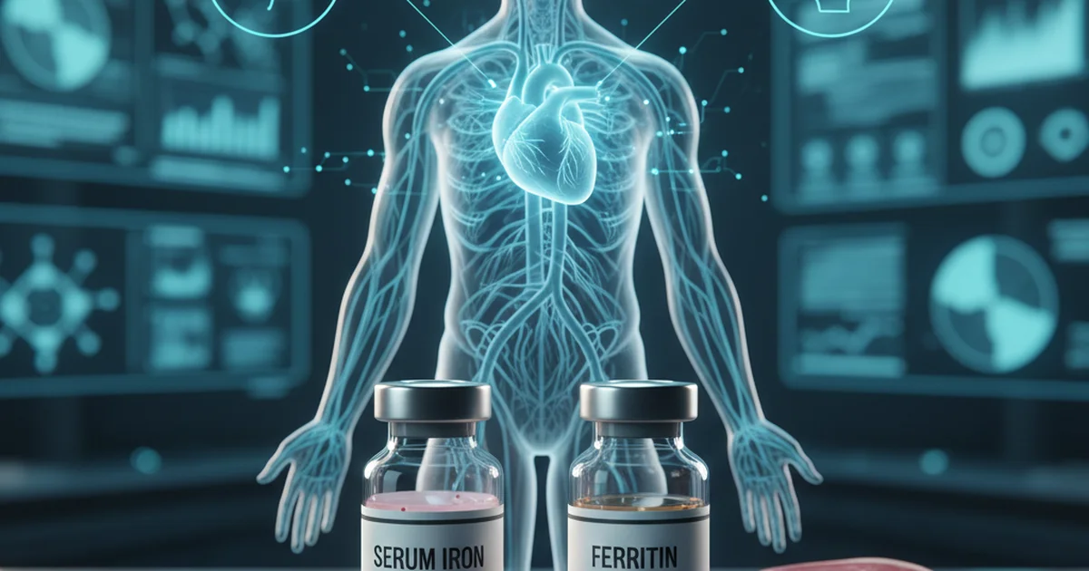
Serum iron measures the amount of circulating iron bound to transferrin, the transport protein that delivers iron to cells. It reflects short‑term iron status and fluctuates throughout the day, typically peaking in the morning and declining after meals. Serum iron is one component of the iron panel, which also includes ferritin (iron storage), total iron‑binding capacity (TIBC), and transferrin saturation. While ferritin is the most reliable indicator of total body iron stores, serum iron is essential for detecting acute changes, monitoring therapy, and guiding supplementation.
Reference Ranges
| Population | Normal Range | Units | Notes |
|---|---|---|---|
| Adult Men (18–65 yr) | 65–176 | µg/dL | Slightly higher due to larger muscle mass |
| Adult Women (18–50 yr) | 50–150 | µg/dL | Lower because of menstrual losses |
| Adult Women (≥ 51 yr) | 65–176 | µg/dL | Post‑menopausal levels converge with men |
| Children (1–12 yr) | 50–120 | µg/dL | Age‑dependent; infants have higher needs |
| Pregnant Women (any trimester) | 30–120 | µg/dL | Physiologic dilution lowers serum iron |
| Elderly (> 65 yr) | 50–150 | µg/dL | May be reduced by chronic disease |
Values may vary slightly by laboratory methodology; always interpret results in the context of the full iron panel and clinical picture.
Causes of Low Serum Iron
Low serum iron can arise from three broad categories: decreased intake/absorption, increased loss, and altered distribution. Understanding the underlying mechanism is crucial for targeted treatment.
1. Inadequate Dietary Intake
- Restrictive diets (vegan, vegetarian, low‑meat) that limit heme iron sources.
- Caloric restriction or eating disorders that reduce overall nutrient consumption.
- Economic or food‑insecurity factors that limit access to iron‑rich foods.
2. Impaired Absorption
- Celiac disease, inflammatory bowel disease, or chronic gastritis that damage the duodenal mucosa where iron is absorbed.
- Helicobacter pylori infection causing chronic gastritis and reduced gastric acidity.
- Medications such as proton‑pump inhibitors (PPIs) and antacids that raise gastric pH, impairing the conversion of ferric (Fe³⁺) to ferrous (Fe²⁺) iron.
- Surgical resections (e.g., bariatric surgery, gastric bypass) that bypass the primary absorption site.
3. Chronic Blood Loss
- Menstrual bleeding (heavy or prolonged menses).
- Gastrointestinal bleeding from ulcers, hemorrhoids, colorectal cancer, or angiodysplasia.
- Parasitic infections (hookworm) in endemic regions.
- Frequent blood donation or therapeutic phlebotomy.
4. Increased Physiologic Demand
- Pregnancy (expansion of maternal blood volume and fetal growth).
- Rapid growth periods (infancy, adolescence).
- Endurance training leading to micro‑hematuria or sweat losses.
5. Redistribution or Sequestration
- Inflammatory states (chronic infection, autoimmune disease) trigger hepcidin production, which blocks iron release from macrophages and reduces intestinal absorption. This “functional iron deficiency” can present with low serum iron despite normal or high ferritin.
Symptoms of Low Serum Iron
When serum iron falls, tissues receive insufficient oxygen, producing a recognizable cluster of signs and symptoms:
- Fatigue, weakness, and reduced exercise tolerance – the most common complaints.
- Pallor of the skin, conjunctivae, and nail beds.
- Shortness of breath on exertion.
- Dizziness or light‑headedness, especially when standing quickly.
- Cold extremities due to peripheral vasoconstriction.
- Headaches and difficulty concentrating (“brain fog”).
- Restless legs syndrome and pica (craving non‑food substances) in severe cases.
- Glossitis (smooth, sore tongue) and cheilosis (cracked corners of the mouth).
Because these manifestations overlap with many other conditions, laboratory confirmation is essential before initiating treatment.
Diagnosing Low Serum Iron
A comprehensive iron panel should include:
- Serum iron – reflects circulating iron at the time of draw.
- Ferritin – gauges iron stores; low ferritin (< 30 ng/mL) confirms deficiency.
- Total iron‑binding capacity (TIBC) – rises when iron stores are depleted.
- Transferrin saturation – calculated as (serum iron ÷ TIBC) × 100; values < 20 % suggest deficiency.
Best practice: Obtain blood samples in the morning, fasting, and avoid recent iron supplementation for at least 24 hours to reduce variability.
Dietary Sources of Iron
Heme Iron (Highly Bioavailable)
| Food | Approximate Iron Content (mg per 100 g) |
|---|---|
| Beef liver | 6.5 |
| Chicken liver | 9.0 |
| Lean beef (sirloin) | 2.6 |
| Pork (loin) | 1.0 |
| Lamb (ground) | 1.8 |
| Fish (sardines, canned in oil) | 2.5 |
| Turkey (dark meat) | 2.3 |
Heme iron absorption rates range from 15–35 % and are less affected by dietary inhibitors.
Non‑Heme Iron (Plant‑Based)
| Food | Approximate Iron Content (mg per 100 g) |
|---|---|
| Lentils (cooked) | 3.3 |
| Chickpeas (cooked) | 2.9 |
| Tofu (firm) | 2.7 |
| Spinach (cooked) | 3.6 |
| Pumpkin seeds | 8.8 |
| Quinoa (cooked) | 1.5 |
| Fortified breakfast cereals | 4–18 (varies) |
Non‑heme iron absorption is typically 2–20 % and can be significantly enhanced or hindered by other dietary components.
Maximizing Iron Bioavailability
Enhancers
- Vitamin C (ascorbic acid): Reduces ferric to ferrous iron, boosting absorption by up to 3‑fold. Pair iron‑rich foods with citrus fruits, bell peppers, strawberries, or tomatoes.
- Meat, fish, poultry (the “meat factor”): Peptides and amino acids from animal protein improve non‑heme iron uptake.
- Fermented foods: Lactic‑acid bacteria can lower pH and release bound iron.
Inhibitors
- Phytates (found in whole grains, legumes, nuts) bind iron; soaking, sprouting, or fermenting reduces phytate levels.
- Polyphenols (tea, coffee, red wine) form insoluble complexes; consume these beverages ≥ 1 hour apart from iron meals.
- Calcium (dairy products, supplements) competitively inhibits iron absorption; separate calcium intake from iron‑rich meals.
- Soy proteins (especially in soy milk) can modestly reduce absorption; choose fortified soy products with added vitamin C.
Practical Meal‑Planning Tips
- Combine a heme source (e.g., a small portion of lean beef) with a vitamin C‑rich side (e.g., bell‑pepper salad).
- Add citrus juice to cooked greens (spinach sautéed with lemon).
- Use cast‑iron cookware; up to 5 mg of iron can leach into foods during cooking, especially with acidic dishes.
- Soak beans and grains overnight, discard the soaking water, and rinse before cooking to lower phytate content.
Supplementation: When Food Isn’t Enough
Indications
- Confirmed iron deficiency anemia (low ferritin, low serum iron, low transferrin saturation).
- Chronic blood loss where dietary intake cannot keep pace.
- Malabsorption syndromes unresponsive to diet alone.
- Pregnancy, when oral supplementation is recommended to meet increased demands.
Forms of Iron Supplements
| Form | Elemental Iron (mg per 100 mg) | Absorption | Common Side Effects |
|---|---|---|---|
| Ferrous sulfate | ~20% | Good | Constipation, nausea, dark stools |
| Ferrous gluconate | ~12% | Moderate | Similar but milder GI upset |
| Ferrous fumarate | ~33% | High | Same GI profile |
| Carbonyl iron | ~20% | Slow, steady release | Lower GI irritation |
| Heme iron polypeptide | ~20% | Highly bioavailable, less dependent on pH | Minimal GI side effects |
| Liquid iron (e.g., ferrous sulfate drops) | Variable | Good for children or those with dysphagia | May cause taste issues |
Key point: Elemental iron is the actual amount of iron available for absorption; dosing is expressed in elemental iron, not the weight of the compound.
Dosing Guidelines
- Adults with deficiency: 100–200 mg elemental iron daily in divided doses (e.g., 2 × 50 mg).
- Pregnant women: 27 mg elemental iron per day (often provided in prenatal vitamins).
- Children (6 mo–12 yr): 3–6 mg/kg elemental iron per day, not exceeding 40 mg per dose.
Administration tips to improve tolerance and absorption:
- Take on an empty stomach (30 minutes before meals) for maximal absorption.
- If gastrointestinal upset occurs, take with a small amount of food or switch to a formulation with lower elemental iron (e.g., ferrous gluconate).
- Avoid calcium, antacids, tea, coffee, or high‑phytate foods within 2 hours of the dose.
- Vitamin C (e.g., a glass of orange juice) taken simultaneously can enhance absorption.
- Re‑evaluate iron status after 4–6 weeks; once ferritin normalizes, reduce the dose to a maintenance level (often 30–60 mg elemental iron weekly) to prevent overload.
Risks of Excess Iron
- Gastrointestinal irritation and constipation.
- Iron overload (hemochromatosis) in genetically predisposed individuals; chronic excess can damage liver, heart, and endocrine organs.
- Interaction with certain antibiotics (e.g., tetracyclines, fluoroquinolones) reducing their effectiveness.
Never self‑prescribe high‑dose iron without laboratory confirmation and medical supervision.
Lifestyle and Medical Management
Dietary Adjustments
- Plan iron‑rich meals at least twice daily, incorporating both heme and non‑heme sources.
- Boost vitamin C intake with fruits, vegetables, or a supplement (250–500 mg daily).
- Limit inhibitors around iron‑rich meals; schedule coffee/tea and calcium‑rich foods at different times.
Address Underlying Causes
- Screen for gastrointestinal bleeding (e.g., fecal occult blood test) if iron deficiency is unexplained.
- Test for celiac disease or H. pylori infection when malabsorption is suspected.
- Review medications that may impair absorption (PPIs, antacids) and discuss alternatives with a clinician.
Monitoring
- Baseline labs: serum iron, ferritin, TIBC, transferrin saturation, complete blood count.
- Follow‑up every 4–8 weeks during treatment; adjust dose based on ferritin trends.
- Long‑term maintenance: once ferritin reaches > 100 ng/mL, reduce supplementation to a maintenance schedule or rely on diet alone.
Special Populations
- Pregnant women: prioritize prenatal vitamins with iron and folic acid; monitor hemoglobin each trimester.
- Athletes: high training volumes may increase iron loss through sweat and hemolysis; consider periodic screening.
- Elderly: chronic inflammation can mask deficiency; evaluate hepcidin levels or use ferritin in conjunction with CRP.
Summary
Low serum iron is a multifactorial condition that can stem from inadequate intake, impaired absorption, chronic blood loss, increased physiological demand, or inflammatory sequestration. Clinical manifestations range from subtle fatigue to overt anemia, underscoring the need for accurate laboratory assessment. Management hinges on three pillars:
- Optimizing dietary iron through strategic food combinations and minimizing inhibitors.
- Targeted supplementation when diet alone cannot meet needs, with careful attention to dosing, formulation, and side‑effect mitigation.
- Addressing root causes—whether gastrointestinal pathology, medication effects, or chronic disease—while monitoring labs to avoid both deficiency and overload.
By integrating evidence‑based nutrition, lifestyle modifications, and appropriate medical oversight, individuals can restore healthy serum iron levels, improve energy, and prevent long‑term complications associated with iron deficiency.
Frequently Asked Questions
What is the most common cause of abnormal Iron (Serum) levels?
The leading cause of low serum iron is dietary insufficiency combined with chronic blood loss, most frequently due to heavy menstrual bleeding in premenopausal women or gastrointestinal bleeding in older adults. In many cases, a combination of modest dietary gaps and a hidden source of loss (e.g., ulcer, hemorrhoids) drives the deficiency.
How often should I get my Iron (Serum) tested?
- If you have known deficiency or are on iron therapy: check serum iron, ferritin, and transferrin saturation every 4–8 weeks until levels normalize.
- Routine screening: for healthy adults without risk factors, a complete blood count (CBC) every 2–3 years is sufficient; targeted iron panels are ordered if anemia or symptoms appear.
- High‑risk groups (pregnant women, athletes, chronic disease patients) should have iron status evaluated each trimester or annually, respectively, or sooner if symptoms develop.
Can lifestyle changes improve my Iron (Serum) levels?
Absolutely. Consistent dietary strategies—such as pairing iron‑rich foods with vitamin C, reducing intake of iron inhibitors around meals, and using cast‑iron cookware—can raise absorption significantly. Additionally, addressing modifiable risk factors (e.g., treating H. pylori infection, adjusting medications that raise gastric pH, managing heavy menstrual flow) and regular physical activity that improves overall circulation also support healthier iron metabolism. When combined with appropriate supplementation, lifestyle modifications often normalize serum iron without the need for high‑dose therapy.
Medical Disclaimer
This article is for educational purposes only. Always consult a healthcare professional.