Low TIBC Levels: Causes and Clinical Meaning
Reference Ranges
| Population | Normal Range | Units | Notes |
|---|---|---|---|
| Adult Men | 250‑450 | µg/dL | Values can vary slightly by laboratory |
| Adult Women | 250‑450 | µg/dL | Slightly lower in pre‑menopausal women |
| Pregnant Women | 200‑400 | µg/dL | Hormonal changes lower TIBC modestly |
| Children 1‑12 yr | 260‑500 | µg/dL | Age‑dependent; higher in early childhood |
| Adolescents 13‑18 yr | 250‑460 | µg/dL | Pubertal surge in iron demand |
| Elderly (>65 yr) | 230‑430 | µg/dL | Decline in hepatic synthesis may occur |
Values are presented as typical laboratory reference intervals; individual labs may report slightly different cut‑offs.
Introduction
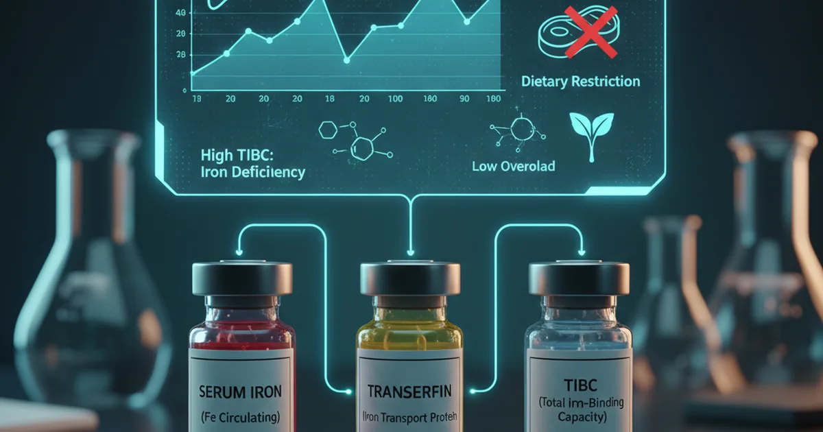
Total Iron‑Binding Capacity (TIBC) is a laboratory measurement that reflects the maximum amount of iron that can be bound by transferrin, the principal iron‑transport protein in plasma. While the test is often ordered alongside serum iron and ferritin to evaluate iron status, clinicians sometimes encounter a low TIBC result and wonder about its significance. Low TIBC is less common than a high TIBC, yet it can be a clue to several metabolic, inflammatory, and nutritional conditions. This article reviews the physiology behind TIBC, explores the causes of a reduced capacity, interprets the clinical meaning, and provides evidence‑based dietary and supplemental strategies to support optimal iron metabolism.
Physiology of Iron and TIBC
What TIBC Measures
- Transferrin saturation is calculated as (Serum Iron ÷ TIBC) × 100.
- TIBC estimates the total binding sites available on circulating transferrin molecules.
- Each transferrin molecule can bind up to two ferric (Fe³⁺) ions; the concentration of transferrin therefore dictates the TIBC value.
Regulation of Transferrin Production
- Hepatocytes synthesize transferrin; production is up‑regulated when iron stores are low (to increase transport capacity) and down‑regulated in states of iron excess or inflammation.
- Cytokines such as interleukin‑6 (IL‑6) suppress transferrin gene expression, contributing to lower TIBC during acute‑phase reactions.
Relationship to Other Iron Indices
| Parameter | High Level Indicates | Low Level Indicates |
|---|---|---|
| Serum Iron | Iron overload, hemolysis | Iron deficiency, chronic blood loss |
| Ferritin | Iron overload, inflammation | Iron deficiency (when low) |
| Transferrin (TIBC) | Iron deficiency, pregnancy | Inflammation, malnutrition, liver disease |
| Transferrin Saturation | Iron overload | Iron deficiency (when low) |
Understanding how TIBC fits within this panel helps clinicians differentiate true iron deficiency from anemia of chronic disease, malnutrition, or hepatic dysfunction.
Causes of Low TIBC
1. Inflammatory and Chronic Disease States
- Anemia of chronic disease (ACD): Elevated IL‑6 and hepcidin reduce transferrin synthesis, leading to low TIBC despite normal or low serum iron.
- Infections (bacterial, viral, fungal) trigger an acute‑phase response that suppresses hepatic production of transferrin.
2. Malnutrition and Protein‑Calorie Deficiency
- Transferrin is a negative acute‑phase protein whose levels fall when dietary protein intake is insufficient.
- Severe caloric restriction, eating disorders, or chronic gastrointestinal malabsorption (e.g., celiac disease) can lower TIBC.
3. Liver Disease
- The liver is the primary site of transferrin synthesis. Cirrhosis, hepatitis, or hepatic steatosis diminish hepatic synthetic function, reducing TIBC.
- Alcoholic liver disease often presents with a pattern of low TIBC, low albumin, and elevated gamma‑glutamyl transferase.
4. Nephrotic Syndrome
- Loss of plasma proteins in the urine includes transferrin, decreasing circulating levels and thus TIBC.
5. Genetic Disorders
- Congenital transferrin deficiency (rare) results from mutations in the TF gene, producing chronically low TIBC.
6. Iron Overload States
- In conditions such as hereditary hemochromatosis, excess iron down‑regulates transferrin synthesis, modestly lowering TIBC. However, the primary laboratory hallmark is a high transferrin saturation, not a markedly low TIBC.
7. Medications and Hormonal Influences
- Oral contraceptives and estrogen therapy can increase hepatic protein synthesis, sometimes raising TIBC; conversely, glucocorticoids may suppress it.
- Chemotherapeutic agents that cause hepatic injury can indirectly lower TIBC.
Clinical Meaning of a Low TIBC
When a low TIBC is identified, the clinician should consider the entire iron panel and the patient’s clinical context.
Anemia of Chronic Disease
- Pattern: Low/normal serum iron, low TIBC, normal‑to‑high ferritin, low transferrin saturation.
- Interpretation: Inflammation sequesters iron within macrophages; the body reduces iron availability to limit pathogen growth.
Nutritional Deficiency
- Pattern: Low serum iron, low TIBC, low ferritin, low transferrin saturation.
- Interpretation: Protein‑calorie malnutrition reduces transferrin production; iron stores may also be depleted.
Liver Dysfunction
- Pattern: Low TIBC with low albumin, elevated bilirubin, and abnormal liver enzymes.
- Interpretation: Impaired hepatic synthesis of transferrin reflects broader synthetic failure.
Nephrotic Syndrome
- Pattern: Low TIBC, hypoalbuminemia, proteinuria >3.5 g/24 h.
- Interpretation: Urinary loss of transferrin reduces circulating levels.
Combined States
- Many patients, especially the elderly, may have overlapping chronic inflammation and mild malnutrition, producing a “mixed” iron‑status picture.
Key clinical takeaway: A low TIBC alone does not diagnose iron deficiency; it signals that the body’s capacity to transport iron is reduced, most often due to inflammation or impaired protein synthesis.
Dietary Sources and Bioavailability of Iron
While TIBC reflects protein synthesis rather than dietary iron directly, the availability of iron in the diet influences the body’s demand for transferrin. Adequate intake of high‑quality protein supports transferrin production, and balanced iron intake prevents secondary alterations in TIBC.
Heme Iron (Highly Bioavailable)
| Food Source | Approx. Heme Iron (mg/100 g) |
|---|---|
| Beef liver | 6.2 |
| Chicken thigh (dark) | 2.7 |
| Pork loin | 2.5 |
| Salmon | 0.8 |
- Absorption rate: 15‑35 % of ingested heme iron is absorbed, largely unaffected by dietary inhibitors.
Non‑Heme Iron (Variable Bioavailability)
| Food Source | Approx. Non‑Heme Iron (mg/100 g) |
|---|---|
| Lentils (cooked) | 3.3 |
| Spinach (cooked) | 2.7 |
| Fortified breakfast cereal | 4.5–18 (depends on brand) |
| Pumpkin seeds | 3.3 |
- Absorption rate: 2‑20 % and highly influenced by enhancers (vitamin C, meat factor) and inhibitors (phytates, polyphenols, calcium).
Protein as a Cofactor for Transferrin Synthesis
- High‑quality animal proteins (eggs, dairy, lean meats) provide essential amino acids necessary for hepatic transferrin synthesis.
- Plant proteins can also support synthesis when intake is adequate and combined with complementary amino acid profiles (e.g., legumes + grains).
Micronutrients that Modify Iron Metabolism
| Nutrient | Effect on Iron Absorption/Utilization |
|---|---|
| Vitamin C | Reduces ferric to ferrous form, boosting non‑heme iron absorption |
| Vitamin A | Enhances mobilization of iron from stores |
| Copper | Required for ceruloplasmin, which oxidizes Fe²⁺ to Fe³⁺ for binding to transferrin |
| Zinc | Excess zinc can compete with iron for absorption sites |
Ensuring a diet rich in these cofactors helps maintain efficient iron transport and may indirectly support normal TIBC values.
Supplementation Strategies When TIBC Is Low
When to Supplement
- Documented iron deficiency (low ferritin, low serum iron) and low TIBC due to malnutrition or protein deficiency.
- Chronic blood loss (e.g., gastrointestinal bleeding) where iron stores are exhausted.
Do not supplement solely on the basis of a low TIBC; inappropriate iron can exacerbate oxidative stress, especially in inflammatory states.
Choice of Iron Form
| Form | Typical Elemental Iron (mg) | Absorption Characteristics |
|---|---|---|
| Ferrous sulfate | 20 % (e.g., 325 mg tablet = 65 mg elemental) | Highest bioavailability; may cause GI upset |
| Ferrous gluconate | 12 % (e.g., 300 mg tablet = 36 mg elemental) | Better tolerated, slightly lower absorption |
| Ferrous fumarate | 33 % (e.g., 200 mg tablet = 66 mg elemental) | High absorption, moderate tolerance |
| Iron polysaccharide complex | 20‑30 % | Minimal GI irritation, slower release |
| Heme iron polypeptide | 10‑15 % | Not affected by dietary inhibitors |
Adjunctive Nutrients for Optimal Absorption
- Vitamin C: 100–200 mg taken with iron increases non‑heme iron absorption by up to 2‑fold.
- Copper: 1–2 mg daily when long‑term iron therapy is used, to prevent secondary copper deficiency.
Dosing Recommendations
- Mild deficiency: 60–120 mg elemental iron daily, divided into two doses to reduce gastrointestinal side effects.
- Severe deficiency or rapid repletion: 150–200 mg elemental iron daily for a limited period (4–6 weeks), then taper.
Monitoring
- Re‑check serum ferritin and hemoglobin after 4–6 weeks of therapy.
- TIBC typically normalizes as iron stores are replenished and hepatic protein synthesis recovers; however, in chronic inflammation it may remain low despite iron repletion.
When Supplementation Is Contraindicated
- Active infection or uncontrolled inflammation (risk of feeding pathogens).
- Hemochromatosis or other iron‑overload disorders.
- Anemia of chronic disease without proven iron deficiency; focus on treating the underlying condition rather than iron loading.
Practical Dietary Recommendations to Support Normal TIBC
Prioritize high‑quality protein
- Aim for 1.0–1.2 g/kg body weight per day (e.g., lean meats, fish, eggs, dairy, soy).
- For vegetarians, combine legumes with grains at each meal to ensure a complete amino‑acid profile.
Include heme iron sources 2–3 times per week to boost iron status without relying on high-dose supplements.
Pair non‑heme iron foods with vitamin C
- Example: Add bell pepper slices to lentil soup, or drizzle lemon juice over spinach salad.
Limit inhibitors around iron‑rich meals
- Avoid tea, coffee, and high‑calcium foods (milk, cheese) within 1 hour of iron intake.
Ensure adequate copper intake
- Include nuts, seeds, shellfish, and whole grains; a typical diet provides 0.9–1.3 mg copper daily.
Address underlying inflammation
- Adopt anti‑inflammatory dietary patterns (Mediterranean style: olive oil, fatty fish, fruits, vegetables, whole grains).
- Weight management, smoking cessation, and regular physical activity can lower systemic IL‑6, indirectly supporting transferrin synthesis.
Consider fortified foods if dietary intake is insufficient
- Breakfast cereals fortified with iron and vitamin C are an efficient way to increase bioavailable iron.
Management of Low TIBC in Specific Clinical Scenarios
| Clinical Situation | Primary Intervention | Role of Diet/Supplementation |
|---|---|---|
| Anemia of chronic disease with low TIBC | Treat underlying inflammation (e.g., disease‑modifying agents, infection control) | Optimize protein intake; avoid unnecessary iron supplementation |
| Malnutrition‑related low TIBC | Nutritional rehabilitation (high‑protein, calorie‑dense meals) | Provide iron‑rich foods once protein status improves; monitor ferritin |
| Chronic liver disease | Manage liver etiology (antiviral therapy, alcohol cessation) | Ensure adequate protein (preferably vegetable‑based if hepatic encephalopathy risk) |
| Nephrotic syndrome | Treat underlying renal disease; replace lost proteins | Oral iron may be needed if serum iron falls; avoid excessive sodium |
| Iron‑deficiency anemia with low TIBC | Oral iron therapy + dietary counseling | Pair iron tablets with vitamin C; space from calcium‑rich meals |
Summary
- Low TIBC is a laboratory clue that the body’s capacity to transport iron is reduced, most commonly due to inflammation, protein‑calorie malnutrition, or liver disease.
- Interpretation requires a full iron panel (serum iron, ferritin, transferrin saturation) and a thorough clinical assessment.
- Dietary protein is essential for hepatic transferrin synthesis; high‑quality animal or complementary plant proteins support normal TIBC.
- Iron‑rich foods (heme and non‑heme) combined with vitamin C enhance absorption, while inhibitors (phytates, polyphenols, calcium) should be spaced away from iron‑containing meals.
- Supplementation is reserved for true iron deficiency or rapid repletion needs; the choice of iron salt, dose, and adjunctive nutrients (vitamin C, copper) influences efficacy and tolerability.
- Addressing the root cause—whether it is chronic inflammation, malnutrition, or hepatic dysfunction—is the cornerstone of normalizing TIBC and overall iron homeostasis.
Frequently Asked Questions
What is the most common cause of abnormal Total Iron Binding Capacity (TIBC) levels?
The most frequent cause of a low TIBC is an inflammatory or chronic disease state, such as anemia of chronic disease, where cytokine‑mediated suppression of hepatic transferrin synthesis reduces the circulating binding capacity.
How often should I get my Total Iron Binding Capacity (TIBC) tested?
For individuals with known iron‑metabolism disorders, clinicians typically repeat the TIBC panel every 3–6 months to monitor trends. In acute inflammatory conditions, testing may be deferred until the inflammatory episode resolves, as TIBC can fluctuate rapidly.
Can lifestyle changes improve my Total Iron Binding Capacity (TIBC) levels?
Yes. Enhancing dietary protein intake, reducing chronic inflammation through a balanced anti‑oxidant‑rich diet, maintaining a healthy weight, limiting alcohol consumption, and managing underlying diseases (e.g., infections, liver disease) can all support hepatic transferrin production and help normalize TIBC.
Medical Disclaimer
This article is for educational purposes only. Always consult a healthcare professional.